Rhizomorphs are generally regarded as structures, where hyphae are connected by various mechanisms and which grow more or less in parallel and more or less closely together over a greater distance. Thus, this definition includes all linearly aggregated hyphal structures (cords, strands) regardless whether or not the hyphae are differentiated to some extent.
Rhizomorph preparations should be done according to the descriptions published by Agerer (1991): Methods in Microbiology 23: 25-73. Only very dark rhizomorphs, or rhizomorphs covered by a lot of strongly light reflecting crystals or soil particles need to be sectioned longitudinally; sections can be made as thick sections by the aid of a cryotome. Often it is sufficient to halve the rhizomorph. The thickness of the sections should be at least double as thick as the thickest hyphae expected.
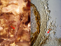
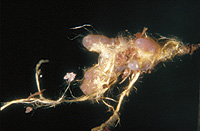
Rhizomorphs must have a natural connection to the mantle cells in order to ensure that they really do belong to the ectomycorrhiza under consideration. Often rhizomorphs of foreign fungi - coloured or colourless ones - can grow on the mantle surface or between the rhizomorphs in question. A rhizomorph is contacting the foreign ectomycorrhiza (A left), and in microscopical view it becomes apparent that there is a delimitation of the rhizomorph from the mantle (A right). In some cases, considerably deviating colours and an obviously secondary cover caused by proximal rhizomorph parts indicate these structures originating from different fungi (B), but microscopical studies have to corroborate such an assumption.
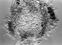
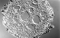
Terminology shown in cross-section (C–D) and in longitudinal (optical) section (E): "vessel-like hyphae" (V) are considerably thicker than all other hyphae and concentrated in central parts, "peripheral hyphae" (pH) form the rim of the rhizomorph and all other hyphae inside of the peripheral hyphae. Those hyaphae which are not vessel-like, are regarded as "central, non-vessel-like hyphae" (cH).
 A |
 B |
 C |
 D |