|
|
|
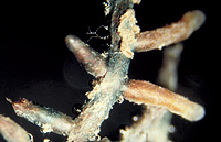
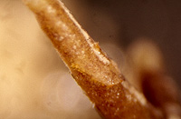
– Enlarged view – |
| • references | |
| Agerer R (1986) Studies on ectomycorrhizae III. Mycorrhizae formed by four fungi in the genera Lactarius and Russula on spruce. Mycotaxon 27: 1-59. Agerer R (1987) Lactarius deterrimus. In Agerer R (ed) Colour Atlas of Ectomycorrhizae, plate 3, Einhorn-Verlag, Schwäbisch Gmünd. Franz F (1994) Ektomykorrhizen der Fichte: Identifizierung, Ultrastruktur und Microelementanalyse (EELS, ESI). Diss Univ Bayreuth. Franz F, Acker G (1995) Rhizomorphs of Picea abies ectomycorrhizae: Ultrastructural aspects and elemental analysis (EELS and ESI) on hyphal inclusions. Nova Hedwigia 60(1-2): 253-267. Münzenberger B, Metzler B, Kottke I, Oberwinkler F (1986) Morphologische und anatomische Charakterisierung der Mykorrhiza Lactarius deterrimus - Picea abies in vitro. Z Mykol 52(2): 407-422. |
|
| • ramification presence-type | |
| monopodial-pinnate | |
| or | monopodial-pyramidal |
| • ramification orders | |
| 0 | Lower value of unspecified range (could be µ-s.d., but not known) |
| 2 | Upper value of unspecified range (could be µ+s.d., but not known) |
| • rhizomorphs as stout, short, conical structures presence-abundance | |
| absent | |
| • rhizomorphs as short mycorrhiza-like outgrowths with blunt tips presence | |
| absent | |
| • rhizomorphs presence | |
| present | |
| • rhizomorphs frequency | |
| infrequent | |
| • exploration type | |
| medium distance smooth | |
| • shape | |
| straight | |
| or | bent |
| or | tortuous |
| • shape {of distal end} | |
| not inflated, cylindric | |
| • length | |
| 0 mm | Lower value of unspecified range (could be µ-s.d., but not known) |
| 2.1 mm | Upper value of unspecified range (could be µ+s.d., but not known) |
| • diameter | |
| 0.32 mm | Lower value of unspecified range (could be µ-s.d., but not known) |
| 0.52 mm | Upper value of unspecified range (could be µ+s.d., but not known) |
| 0.65 mm | Maximum value |
| • colour | |
| orange | |
| • very tip colour | |
| orange | |
| • older parts colour | |
| brown | |
| or | orange |
| or | green |
| • mantle {distinct} surface visibility | |
| present | |
| • mantle transparency | |
| not transparent | |
| • mantle secreted latex {after scratching mantle} presence | |
| present | |
| • mantle secreted latex {after scratching mantle} colour-habit | |
| orange | |
| • mantle carbonizing presence | |
| absent | |
| • mantle surface {in general} habit | |
| smooth | |
| • cross-section shape | |
| round or roundish | |
| • colour | |
| orange | |
| or | green |
| • ramification kind-frequency | |
| infrequently, at restricted points | |
| • margin habit | |
| smooth | |
| • dimorphism presence | |
| absent | |
| • presence | |
| absent | |
| • emanating elements presence-type | |
| rhizomorphs | |
| • presence | |
| present | |
| • location | |
| middle mantle layer | |
| or | inner mantle layer |
| or | rhizomorphs |
| • shape | |
| straight and even | |
| • cell diameter | |
| 3 µm | Lower value of unspecified range (could be µ-s.d., but not known) |
| 6 µm | Upper value of unspecified range (could be µ+s.d., but not known) |
| • matrix presence | |
| present | |
| • matrix location | |
| outer mantle layer {apart from tip} | |
| • organisation | |
| plectenchymatous | |
| • mantle type | |
| hyphae rather irregularly arranged and no special pattern discernible (type B) | |
| • matrix kind | |
| not gelatinous | |
| • hyphal system kind | |
| undifferentiated | |
| • septa clamps presence | |
| absent | |
| • cell pigment location-colour | |
| plasmatically yellowish | |
| and | vacuolarly yellowish |
| • cell diameter | |
| 2 µm | Lower value of unspecified range (could be µ-s.d., but not known) |
| 3 µm | Upper value of unspecified range (could be µ+s.d., but not known) |
| 3.5 µm | Maximum value |
| • cell wall thickness | |
| 0.5 µm | Lower value of unspecified range (could be µ-s.d., but not known) |
| 1 µm | Upper value of unspecified range (could be µ+s.d., but not known) |
| • cell wall surface habit | |
| smooth | |
| • drops of exuded pigment presence | |
| absent | |
| • organisation | |
| plectenchymatous | |
| • hyphae arrangement | |
| without pattern | |
| • septa clamps presence | |
| absent | |
| • cell diameter | |
| 2 µm | Minimum value |
| 3 µm | Lower value of unspecified range (could be µ-s.d., but not known) |
| 4 µm | Upper value of unspecified range (could be µ+s.d., but not known) |
| 7 µm | Maximum value |
| • cell contents presence-kind | |
| absent | |
| • mantle thickness {apart from tip} | |
| 10 µm | Minimum value |
| 20 µm | Lower value of unspecified range (could be µ-s.d., but not known) |
| 30 µm | Upper value of unspecified range (could be µ+s.d., but not known) |
| 40 µm | Maximum value |
| • mantle different layers presence | |
| not discernable | |
| or | discernable |
| • outer mantle layer organisation | |
| plectenchymatous | |
| • middle mantle layer organisation | |
| plectenchymatous | |
| • inner mantle layer organisation | |
| plectenchymatous | |
| • unlayered mantle hyphae tangentially length | |
| 2 µm | Lower value of unspecified range (could be µ-s.d., but not known) |
| 10 µm | Upper value of unspecified range (could be µ+s.d., but not known) |
| 20 µm | Maximum value |
| • unlayered mantle hyphae radially diameter | |
| 2 µm | Lower value of unspecified range (could be µ-s.d., but not known) |
| 3 µm | Upper value of unspecified range (could be µ+s.d., but not known) |
| 5 µm | Maximum value |
| • presence | |
| present | |
| • rows number | |
| 1 | Lower value of unspecified range (could be µ-s.d., but not known) |
| 2 | Upper value of unspecified range (could be µ+s.d., but not known) |
| 3 | Maximum value |
| • shape | |
| tangentially-oval, -elliptic or -cylindrical, and oriented in parallel to root axis | |
| • tangentially length | |
| 50 µm | Lower value of unspecified range (could be µ-s.d., but not known) |
| 90 µm | Upper value of unspecified range (could be µ+s.d., but not known) |
| 120 µm | Maximum value |
| • radially diameter | |
| 5 µm | Lower value of unspecified range (could be µ-s.d., but not known) |
| 12 µm | Upper value of unspecified range (could be µ+s.d., but not known) |
| • anatomy mantle longitudinal section cortical (epidermal) cells shape | |
| tangentially-oval to -elliptic or -cylindrical, and oriented in parallel to root axis | |
| • anatomy mantle longitudinal section cortical (epidermal) cells tangentially length | |
| 60 µm | Lower value of unspecified range (could be µ-s.d., but not known) |
| 110 µm | Upper value of unspecified range (could be µ+s.d., but not known) |
| • anatomy mantle longitudinal section cortical (epidermal) cells radially diameter | |
| 12 µm | Lower value of unspecified range (could be µ-s.d., but not known) |
| 20 µm | Upper value of unspecified range (could be µ+s.d., but not known) |
| 30 µm | Maximum value |
| • presence | |
| present | |
| • kind | |
| protruding towards endodermis | |
| or | one or half a row of cortical cells adjoining endodermis free of Hartig net |
| • anatomy mantle longitudinal section hyphal cells around tannin cells shape | |
| roundish | |
| • anatomy mantle longitudinal section hyphal rows around tannin cells number | |
| one | |
| or | two |
| • structure {in plan view} | |
| of palmetti type | |
| • mantle different layers presence | |
| not discernible | |
| • outer mantle layer organisation | |
| plectenchymatous | |
| • middle mantle layer organisation | |
| plectenchymatous | |
| • inner mantle layer organisation | |
| plectenchymatous | |
| • unlayered mantle hyphae tangentially length | |
| 2 µm | Lower value of unspecified range (could be µ-s.d., but not known) |
| 10 µm | Upper value of unspecified range (could be µ+s.d., but not known) |
| 15 µm | Maximum value |
| • unlayered mantle hyphae radially diameter | |
| 1 µm | Lower value of unspecified range (could be µ-s.d., but not known) |
| 2.5 µm | Upper value of unspecified range (could be µ+s.d., but not known) |
| • laticifers diameter | |
| 4 µm | Lower value of unspecified range (could be µ-s.d., but not known) |
| 5 µm | Upper value of unspecified range (could be µ+s.d., but not known) |
| • presence | |
| present | |
| • shape | |
| tangentially-oval to tangentially-elliptic | |
| • tangentially length | |
| 30 µm | Lower value of unspecified range (could be µ-s.d., but not known) |
| 40 µm | Upper value of unspecified range (could be µ+s.d., but not known) |
| • radially diameter | |
| 5 µm | Lower value of unspecified range (could be µ-s.d., but not known) |
| 10 µm | Upper value of unspecified range (could be µ+s.d., but not known) |
| • anatomy mantle cross-section cortical (epidermal) cells shape | |
| tangentially-oval to tangentially-elliptic | |
| • anatomy mantle cross-section cortical (epidermal) cells tangentially length | |
| 30 µm | Lower value of unspecified range (could be µ-s.d., but not known) |
| 60 µm | Upper value of unspecified range (could be µ+s.d., but not known) |
| • anatomy mantle cross-section cortical (epidermal) cells radially diameter | |
| 10 µm | Minimum value |
| 15 µm | Lower value of unspecified range (could be µ-s.d., but not known) |
| 20 µm | Upper value of unspecified range (could be µ+s.d., but not known) |
| 30 µm | Maximum value |
| • intrahyphal hyphae presence | |
| present | |
| • anastomoses location | |
| not specified | |
| • shape | |
| not striking | |
| • cell pigment location-colour | |
| absent | |
| • drops of exuded pigment presence | |
| absent | |
| • clamps presence | |
| absent | |
| • anatomy emanating elements emanating hyphae cell diameter | |
| 2.5 µm | Mean (= average) |
| • anatomy emanating elements emanating hyphae cell wall surface habit | |
| without lens-shaped appositions | |
| or | without spindle-shaped appositions |
| • anatomy emanating elements emanating hyphae cell wall thickness | |
| 0.5 µm | Mean (= average) |
| • type | |
| undifferentiated; margins rather smooth; hyphae compactly arranged and of uniform diameter (type B) |
|
| or | differentiated; some hyphae very thick, which appear randomly distributed, pores of septa somtimes enlarged (type D) |
| • internal nodia presence | |
| absent | |
| • a "ball" of intertwined, ramified, thin hyphae presence | |
| absent | |
| • presence | |
| absent | |
| • {of ectomycorrhiza former} presence | |
| absent | |
| • {of foreign origin} presence | |
| absent | |
| • number {per cell} | |
| 2 | Lower value of unspecified range (could be µ-s.d., but not known) |
| 4 | Upper value of unspecified range (could be µ+s.d., but not known) |
| • presence | |
| absent | |
| • septal pores type | |
| as dolipores with perforated parenthesome | |
| • {occurence in} humus type | |
| raw humus | |
| • geographic occurrence continent | |
| Europe | |
| • knowledge about association with foreign fruitbodies presence | |
| unknown | |
| • plant family | |
| Pinaceae | |
| • plant genus | |
| Picea | |
| • plant habitat kind | |
| forests, woods | |
| • family | |
| Russulaceae | |
| • subgenus-section | |
| Lactarius sect. Dapetes | |
| • fruitbodies growth habit | |
| epigeous | |
| or | pileate-lamellate |
| • public notes | |
| Laticifers without septa; autofluorescence of mantle in section with UV-filter blue and laticifers distinctly blue, with blue-filter greenish and laticifers distinctly green; mantle in sulfo-vanillin in general red-brown and laticifers with dark granules, in KOH ochre-orange, especially laticifers, in brillant-cresyl-blue bluish-green and laticifers conspicuously so, in toluidin-blue violet-blue, in cotton-blue slightly blue, in a-naphthol no reaction, in NH4OH ochre-orange and particularly laticifers, in chlorazol-black slightly bluish green, in erythrosin reddish, in fast-green green; nuclei lying close togehter. | |