|
|
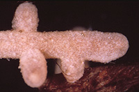
– Enlarged view – |
| • references | |
| Brand F (1988) Fagirhiza granulosa. In Agerer R (ed) Colour Atlas of Ectomycorrhizae, plate 16, Einhorn-Verlag, Schwäbisch Gmünd. Brand F (1991) Ektomykorrhizen an Fagus sylvatica. Charakterisierung und Identifizierung, ökologische Kennzeichnung und unsterile Kultivierung. Libri Botanici 2: 1-229. Brand F, Agerer R (1987) Studien an Ektomykorrhizen XIII. Drei Häufige Ektomykorrhizen der Buche (Fagus sylvatica L.). Charakterisierung und unsterile Kultivierung von Buchenektomykorrhizen. Sydowia 40: 1-37. - Beenken L (2004) Die Gattung Russula. Untersuchungen zu ihrer Systematik anhand von Ektomykorrhizen. Dissertation, Universität München. http://edoc.ub.uni-muenchen.de/archive/00003175/01/Beenken_Ludwig.pdf |
|
| • length | |
| 0 mm | Lower value of unspecified range (could be µ-s.d., but not known) |
| 3 mm | Upper value of unspecified range (could be µ+s.d., but not known) |
| • ramification presence-type | |
| monopodial-pyramidal | |
| or | coralloid |
| • ramification orders | |
| 0 | Lower value of unspecified range (could be µ-s.d., but not known) |
| 1 | Upper value of unspecified range (could be µ+s.d., but not known) |
| • main axis diameter | |
| 0.3 mm | Mean (= average) |
| • rhizomorphs as stout, short, conical structures presence-abundance | |
| absent | |
| • rhizomorphs as short mycorrhiza-like outgrowths with blunt tips presence | |
| absent | |
| • rhizomorphs presence | |
| absent | |
| • shape | |
| straight | |
| or | bent |
| • shape {of distal end} | |
| not inflated, cylindric | |
| • length | |
| 0 mm | Lower value of unspecified range (could be µ-s.d., but not known) |
| 1.5 mm | Upper value of unspecified range (could be µ+s.d., but not known) |
| • diameter | |
| 0.3 mm | Lower value of unspecified range (could be µ-s.d., but not known) |
| 0.4 mm | Upper value of unspecified range (could be µ+s.d., but not known) |
| • colour | |
| brown | |
| or | green |
| • very tip colour | |
| brown | |
| • mantle cortical cells visibility | |
| not visible | |
| • mantle {distinct} surface visibility | |
| present | |
| • mantle transparency | |
| not transparent | |
| • mantle laticifers visibility | |
| absent | |
| • mantle dots presence-colour | |
| absent | |
| • mantle carbonizing presence | |
| absent | |
| • mantle surface {in general} habit | |
| not smooth | |
| • mantle surface {in detail} kind | |
| densely grainy or warty | |
| • emanating hyphae presence | |
| present | |
| • emanating hyphae abundance | |
| infrequent | |
| • presence | |
| absent | |
| • emanating elements presence-type | |
| cystidia | |
| • emanating elements cystidia location | |
| on outer mantle layer | |
| • presence | |
| absent | |
| • anatomy entire mycorrhiza laticiferous solitary (gloeoplerous) cells presence | |
| present | |
| • organisation | |
| pseudoparenchymatous | |
| • organisation {if pseudoparenchymatous} cell shape | |
| angular | |
| • mantle type | |
| angular cells, bearing mounds of roundish cells (type K) | |
| and | some cells containing droplets, staining in sulpho-vanillin; shape variable (type N) |
| • hyphal net clamps presence | |
| absent | |
| • mound cells granular contents | |
| absent | |
| • pores between cells presence | |
| absent | |
| • septa thickness {relative to cell walls} | |
| thinner than walls | |
| • septa clamps presence | |
| absent | |
| • cell pigment location-colour | |
| membranaceously brownish | |
| • cell contents presence-kind | |
| droplets, that stain in sulpho-vanillin | |
| • cell diameter | |
| 10 µm | Mean (= average) |
| • cell length | |
| 10 µm | Mean (= average) |
| • cell density | |
| 8 | Mean (= average) |
| • cell wall thickness | |
| 0.2 µm | Mean (= average) |
| • cell wall with globular thickenings | |
| absent | |
| • cell wall surface habit | |
| smooth | |
| • cell wall projections presence | |
| absent | |
| • drops of exuded pigment presence | |
| absent | |
| • organisation | |
| plectenchymatous | |
| • hyphae arrangement | |
| without pattern | |
| • septa clamps presence | |
| absent | |
| • cell diameter | |
| 2 µm | Lower value of unspecified range (could be µ-s.d., but not known) |
| 3 µm | Upper value of unspecified range (could be µ+s.d., but not known) |
| 3.5 µm | Maximum value |
| • cell contents presence-kind | |
| absent | |
| • anatomy mantle outer mantle layer {of ectomycorrhizal tip} hyphae diameter | |
| 3 µm | Lower value of unspecified range (could be µ-s.d., but not known) |
| 5 µm | Upper value of unspecified range (could be µ+s.d., but not known) |
| • anatomy mantle outer mantle layer {of ectomycorrhizal tip} cell density | |
| 17 | Mean (= average) |
| • mantle thickness {apart from tip} | |
| 20 µm | Lower value of unspecified range (could be µ-s.d., but not known) |
| 30 µm | Upper value of unspecified range (could be µ+s.d., but not known) |
| 35 µm | Maximum value |
| • mantle different layers presence | |
| discernable | |
| • outer mantle layer organisation | |
| plectenchymatous | |
| • outer mantle layer hyphae tangentially length | |
| 5 µm | Lower value of unspecified range (could be µ-s.d., but not known) |
| 15 µm | Upper value of unspecified range (could be µ+s.d., but not known) |
| • outer mantle layer hyphae radially diameter | |
| 5 µm | Lower value of unspecified range (could be µ-s.d., but not known) |
| 15 µm | Upper value of unspecified range (could be µ+s.d., but not known) |
| • middle mantle layer organisation | |
| pseudoparenchymatous | |
| • middle mantle layer hyphae tangentially length | |
| 5 µm | Lower value of unspecified range (could be µ-s.d., but not known) |
| 15 µm | Upper value of unspecified range (could be µ+s.d., but not known) |
| • middle mantle layer hyphae radially diameter | |
| 4 µm | Lower value of unspecified range (could be µ-s.d., but not known) |
| 7 µm | Upper value of unspecified range (could be µ+s.d., but not known) |
| • inner mantle layer organisation | |
| plectenchymatous | |
| • inner mantle layer hyphae radially diameter | |
| 3 µm | Mean (= average) |
| 5 µm | Maximum value |
| • presence | |
| absent | |
| • anatomy mantle longitudinal section cortical (epidermal) cells shape | |
| radially-oval to -elliptic, oriented obliquely | |
| • anatomy mantle longitudinal section cortical (epidermal) cells tangentially length | |
| 10 µm | Lower value of unspecified range (could be µ-s.d., but not known) |
| 15 µm | Upper value of unspecified range (could be µ+s.d., but not known) |
| • anatomy mantle longitudinal section cortical (epidermal) cells radially diameter | |
| 30 µm | Lower value of unspecified range (could be µ-s.d., but not known) |
| 50 µm | Upper value of unspecified range (could be µ+s.d., but not known) |
| • presence | |
| present | |
| • kind | |
| periepidermal | |
| or | in two rows, not reaching endodermis |
| • mantle different layers presence | |
| discernible | |
| • outer mantle layer organisation | |
| plectenchymatous | |
| • outer mantle layer hyphae tangentially length | |
| 5 µm | Lower value of unspecified range (could be µ-s.d., but not known) |
| 15 µm | Upper value of unspecified range (could be µ+s.d., but not known) |
| • outer mantle layer hyphae radially diameter | |
| 5 µm | Lower value of unspecified range (could be µ-s.d., but not known) |
| 15 µm | Upper value of unspecified range (could be µ+s.d., but not known) |
| • middle mantle layer organisation | |
| pseudoparenchymatous | |
| • middle mantle layer hyphae tangentially length | |
| 5 µm | Lower value of unspecified range (could be µ-s.d., but not known) |
| 15 µm | Upper value of unspecified range (could be µ+s.d., but not known) |
| • middle mantle layer hyphae radially diameter | |
| 4 µm | Lower value of unspecified range (could be µ-s.d., but not known) |
| 7 µm | Upper value of unspecified range (could be µ+s.d., but not known) |
| • inner mantle layer organisation | |
| plectenchymatous | |
| • inner mantle layer hyphae radially diameter | |
| 3 µm | Mean (= average) |
| 5 µm | Maximum value |
| • presence | |
| absent | |
| • anatomy mantle cross-section cortical (epidermal) cells shape | |
| rectangular | |
| or | radially-oval to -elliptic |
| • anatomy mantle cross-section cortical (epidermal) cells tangentially length | |
| 10 µm | Lower value of unspecified range (could be µ-s.d., but not known) |
| 15 µm | Upper value of unspecified range (could be µ+s.d., but not known) |
| • anatomy mantle cross-section cortical (epidermal) cells radially diameter | |
| 10 µm | Lower value of unspecified range (could be µ-s.d., but not known) |
| 25 µm | Upper value of unspecified range (could be µ+s.d., but not known) |
| • presence | |
| present | |
| • kind | |
| apparently two rows deep | |
| or | in tow rows, not reaching endodermis |
| • anatomy mantle cross-section hyphal cells around cortical (epidermal) cells shape | |
| roundish | |
| or | cylindrical |
| • anatomy mantle cross-section hyphal cells around cortical (epidermal) cells thickness | |
| 2 µm | Lower value of unspecified range (could be µ-s.d., but not known) |
| 3 µm | Upper value of unspecified range (could be µ+s.d., but not known) |
| • lobes width | |
| 1 µm | Minimum value |
| 2 µm | Lower value of unspecified range (could be µ-s.d., but not known) |
| 4 µm | Upper value of unspecified range (could be µ+s.d., but not known) |
| • structure {in plan view} | |
| of palmetti type | |
| • type | |
| fusiform (type F) | |
| • septa presence | |
| absent | |
| or | present |
| • septa kind | |
| absent, with basal clamp only | |
| or | simple |
| • diameter {proximal} | |
| 5 µm | Mean (= average) |
| • length | |
| 10 µm | Lower value of unspecified range (could be µ-s.d., but not known) |
| 20 µm | Upper value of unspecified range (could be µ+s.d., but not known) |
| • cell wall colour | |
| yellowish | |
| or | brownish |
| • cell wall thickness | |
| 0.5 µm | Mean (= average) |
| • cell wall thickness {relative to mantle cells} | |
| similar in thickness | |
| or | thicker |
| • cell wall evenness | |
| even in thickness | |
| • surface habit | |
| warty | |
| • contents presence | |
| present | |
| • contents type | |
| finely granular | |
| • shape | |
| not striking | |
| • cell pigment location-colour | |
| membranaceously brownish | |
| • drops of exuded pigment presence | |
| absent | |
| • side-branches at septum number | |
| one side-branch at septum | |
| • clamps presence | |
| absent | |
| • anatomy emanating elements emanating hyphae cell shape | |
| even | |
| • anatomy emanating elements emanating hyphae cell diameter | |
| 2 µm | Lower value of unspecified range (could be µ-s.d., but not known) |
| 3 µm | Upper value of unspecified range (could be µ+s.d., but not known) |
| • anatomy emanating elements emanating hyphae cell length | |
| 10 µm | Lower value of unspecified range (could be µ-s.d., but not known) |
| 15 µm | Upper value of unspecified range (could be µ+s.d., but not known) |
| • anatomy emanating elements emanating hyphae cell wall surface habit | |
| smooth | |
| or | without lens-shaped appositions |
| or | without spindle-shaped appositions |
| • anatomy emanating elements emanating hyphae cell wall thickness | |
| 0.2 µm | Mean (= average) |
| • anatomy emanating elements emanating hyphae cell wall {apart from tip} evenness | |
| even in thickness | |
| • type | |
| lacking, only emanating hyphae present (type G) |
|
| • presence | |
| absent | |
| • {of ectomycorrhiza former} presence | |
| absent | |
| • {of foreign origin} presence | |
| absent | |
| • reaction with brillant-cresyl-blue presence | |
| absent | |
| • reaction with cotton-blue-lactic-acid presence | |
| absent | |
| • reaction with ethanol 70% presence | |
| present | |
| • reaction with FeSO4 presence | |
| absent | |
| • reaction with KOH 10% presence | |
| present | |
| • reaction with lactic acid presence | |
| absent | |
| • reaction with Melzer's reagent presence | |
| dextrinoid | |
| • reaction with phenole presence | |
| absent | |
| • reaction with ruthenium-red presence | |
| absent | |
| • reaction with sudan III presence | |
| absent | |
| • reaction with sulpho-vanillin presence | |
| absent [contents of gloeoplerous cells greyish blue] | |
| or | present |
| • reaction with toluidin blue presence | |
| present | |
| • substrate | |
| in organic layer | |
| or | in organically enriched mineral horizon |
| • geographic occurrence continent | |
| Europe | |
| • knowledge about association with foreign fruitbodies presence | |
| unknown | |
| • plant family | |
| Fagaceae | |
| • plant genus | |
| Fagus | |
| • plant habitat kind | |
| forests, woods | |
| • family | |
| Russulaceae | |
| • subgenus-section | |
| Russula sect. Ingratae | |
| • subsection | |
| Russula sect. Ingratae subsect. Felleinae | |
| • fruitbodies growth habit | |
| epigeous | |
| or | pileate-lamellate |
| • public notes | |
| Mycorrhizal ends orange-brown or brown-orange; autofluorescence of mantle in section with UV-filter slightly orange-brown to lemon-yellow, inner mantle layers partially whitish, with blue-filter slightly greenish yellow to green-orange; mantle in sulfo-vanillin becoming brownish, in KOH walls darker and becoming red-brown, in lactic acid not bleaching, in toluidin- blue bluish, in ethanol slowly bleaching, in chlorazol-black no reaction. | |