|
|
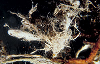
– Enlarged view – |
| • references | |
| Agerer R (1987) Studies on ectomycorrhizae IX. Mycorrhizae formed by Tricholoma sulfureum and T. vaccinum on spruce. Mycotaxon 28: 327-360. Agerer R (1987) Tricholoma sulphureum. In Agerer R (ed) Colour Atlas of Ectomycorrhizae, plate 9, Einhorn-Verlag, Schwäbisch Gmünd. |
|
| • ramification presence-type | |
| monopodial-pinnate | |
| • ramification orders | |
| 0 | Lower value of unspecified range (could be µ-s.d., but not known) |
| 1 | Upper value of unspecified range (could be µ+s.d., but not known) |
| • rhizomorphs as stout, short, conical structures presence-abundance | |
| absent | |
| • rhizomorphs as short mycorrhiza-like outgrowths with blunt tips presence | |
| absent | |
| • rhizomorphs presence | |
| present | |
| • rhizomorphs frequency | |
| abundant | |
| • exploration type | |
| medium distance fringe | |
| • shape | |
| straight | |
| • shape {of distal end} | |
| not inflated, cylindric | |
| • length | |
| 2 mm | Mean (= average) |
| • diameter | |
| 0.3 mm | Mean (= average) |
| 0.33 mm | Maximum value |
| • colour | |
| white | |
| • very tip colour | |
| whitish | |
| • older parts colour | |
| dark brown | |
| or | brown |
| or | ochre, yellowish brown |
| • mantle cortical cells visibility | |
| not visible | |
| • mantle {distinct} surface visibility | |
| present | |
| • mantle transparency | |
| not transparent | |
| • mantle laticifers visibility | |
| absent | |
| • mantle dots presence-colour | |
| absent | |
| • mantle carbonizing presence | |
| absent | |
| • mantle surface {in general} habit | |
| silvery | |
| or | not smooth |
| • mantle surface {in detail} kind | |
| loosely stringy | |
| • emanating hyphae presence | |
| present | |
| • emanating hyphae abundance | |
| abundant | |
| • emanating hyphae distribution | |
| not specifically distributed | |
| • cross-section shape | |
| round or roundish | |
| or | flat |
| • ramification kind-frequency | |
| frequently, at restricted points | |
| • connection to mantle kind | |
| distinct | |
| or | oblique |
| • origin location | |
| proximal | |
| • margin habit | |
| hairy | |
| • dimorphism presence | |
| absent | |
| • presence | |
| absent | |
| • emanating elements presence-type | |
| rhizomorphs | |
| • presence | |
| absent | |
| • organisation | |
| plectenchymatous | |
| • mantle type | |
| ring-like arrangement of hyphal bundles (type A) | |
| • septa clamps presence | |
| absent | |
| • cell shape | |
| cylindric, not constricted at septa | |
| • cell diameter | |
| 3 µm | Lower value of unspecified range (could be µ-s.d., but not known) |
| 4 µm | Upper value of unspecified range (could be µ+s.d., but not known) |
| 5 µm | Maximum value |
| • cell wall thickness | |
| 0.2 µm | Lower value of unspecified range (could be µ-s.d., but not known) |
| 0.5 µm | Upper value of unspecified range (could be µ+s.d., but not known) |
| • drops of exuded pigment presence | |
| absent | |
| • organisation | |
| plectenchymatous | |
| • organisation | |
| plectenchymatous | |
| or | plectenchymatous with pseudoparenchymatous nests of cells |
| • hyphae arrangement | |
| ring-like | |
| • septa clamps presence | |
| absent | |
| • cell diameter | |
| 2 µm | Lower value of unspecified range (could be µ-s.d., but not known) |
| 3 µm | Upper value of unspecified range (could be µ+s.d., but not known) |
| 4 µm | Maximum value |
| • anatomy mantle outer mantle layer {of ectomycorrhizal tip} organisation | |
| like other parts of mantle | |
| • anatomy mantle outer mantle layer {of ectomycorrhizal tip} hyphae diameter | |
| 2 µm | Lower value of unspecified range (could be µ-s.d., but not known) |
| 3.5 µm | Upper value of unspecified range (could be µ+s.d., but not known) |
| • mantle thickness {apart from tip} | |
| 5 µm | Minimum value |
| 10 µm | Mean (= average) |
| 15 µm | Maximum value |
| • mantle thickness {at ectomycorrhizal tip} | |
| 15 µm | Lower value of unspecified range (could be µ-s.d., but not known) |
| 25 µm | Upper value of unspecified range (could be µ+s.d., but not known) |
| • mantle different layers presence | |
| discernable | |
| • outer mantle layer hyphae tangentially length | |
| 3 µm | Minimum value |
| 10 µm | Lower value of unspecified range (could be µ-s.d., but not known) |
| 30 µm | Upper value of unspecified range (could be µ+s.d., but not known) |
| 50 µm | Maximum value |
| • outer mantle layer hyphae radially diameter | |
| 3 µm | Lower value of unspecified range (could be µ-s.d., but not known) |
| 4 µm | Upper value of unspecified range (could be µ+s.d., but not known) |
| • inner mantle layer hyphae tangentially length | |
| 5 µm | Minimum value |
| 10 µm | Lower value of unspecified range (could be µ-s.d., but not known) |
| 20 µm | Upper value of unspecified range (could be µ+s.d., but not known) |
| 30 µm | Maximum value |
| • inner mantle layer hyphae radially diameter | |
| 2.5 µm | Minimum value |
| 3 µm | Lower value of unspecified range (could be µ-s.d., but not known) |
| 4 µm | Upper value of unspecified range (could be µ+s.d., but not known) |
| 5 µm | Maximum value |
| • presence | |
| present | |
| • shape | |
| tangentially-oval to -elliptic or -cylindrical, and oriented obliquely | |
| or | tangentially-oval, -elliptic or -cylindrical, and oriented in parallel to root axis |
| • tangentially length | |
| 18 µm | Minimum value |
| 30 µm | Lower value of unspecified range (could be µ-s.d., but not known) |
| 80 µm | Upper value of unspecified range (could be µ+s.d., but not known) |
| 105 µm | Maximum value |
| • radially diameter | |
| 4 µm | Minimum value |
| 6 µm | Lower value of unspecified range (could be µ-s.d., but not known) |
| 14 µm | Upper value of unspecified range (could be µ+s.d., but not known) |
| 17 µm | Maximum value |
| • mean tangenial length TCt | |
| 56.5 µm | Mean (= average) |
| • mean shape-ratio TCq | |
| 6.7 | Mean (= average) |
| • anatomy mantle longitudinal section cortical (epidermal) cells shape | |
| tangentially-oval to -elliptic or -cylindrical, and oriented obliquely | |
| • anatomy mantle longitudinal section cortical (epidermal) cells tangentially length | |
| 20 µm | Minimum value |
| 35 µm | Lower value of unspecified range (could be µ-s.d., but not known) |
| 90 µm | Upper value of unspecified range (could be µ+s.d., but not known) |
| 102 µm | Maximum value |
| • anatomy mantle longitudinal section cortical (epidermal) cells radially diameter | |
| 8 µm | Minimum value |
| 12 µm | Lower value of unspecified range (could be µ-s.d., but not known) |
| 25 µm | Upper value of unspecified range (could be µ+s.d., but not known) |
| 32 µm | Maximum value |
| • anatomy mantle longitudinal section cortical (epidermal) cells mean tangential length CCt (ECt) | |
| 62.2 µm | Mean (= average) |
| • anatomy mantle longitudinal section cortical (epidermal) cells mean shape-ratio CCq (ECq) | |
| 3.1 | Mean (= average) |
| • mantle different layers presence | |
| discernible | |
| • outer mantle layer organisation | |
| plectenchymatous | |
| • outer mantle layer hyphae tangentially length | |
| 3 µm | Lower value of unspecified range (could be µ-s.d., but not known) |
| 15 µm | Upper value of unspecified range (could be µ+s.d., but not known) |
| 30 µm | Maximum value |
| • outer mantle layer hyphae radially diameter | |
| 2 µm | Lower value of unspecified range (could be µ-s.d., but not known) |
| 4 µm | Upper value of unspecified range (could be µ+s.d., but not known) |
| 5 µm | Maximum value |
| • middle mantle layer organisation | |
| plectenchymatous | |
| • inner mantle layer organisation | |
| plectenchymatous | |
| • inner mantle layer hyphae tangentially length | |
| 3 µm | Lower value of unspecified range (could be µ-s.d., but not known) |
| 7 µm | Upper value of unspecified range (could be µ+s.d., but not known) |
| 15 µm | Maximum value |
| • inner mantle layer hyphae radially diameter | |
| 2 µm | Lower value of unspecified range (could be µ-s.d., but not known) |
| 4 µm | Upper value of unspecified range (could be µ+s.d., but not known) |
| 5 µm | Maximum value |
| • presence | |
| present | |
| • rows number | |
| 1 | Minimum value |
| 2 | Mean (= average) |
| 3 | Maximum value |
| • shape | |
| tangentially-oval to tangentially-elliptic | |
| • tangentially length | |
| 15 µm | Minimum value |
| 25 µm | Lower value of unspecified range (could be µ-s.d., but not known) |
| 50 µm | Upper value of unspecified range (could be µ+s.d., but not known) |
| 60 µm | Maximum value |
| • radially diameter | |
| 4 µm | Minimum value |
| 7 µm | Lower value of unspecified range (could be µ-s.d., but not known) |
| 15 µm | Upper value of unspecified range (could be µ+s.d., but not known) |
| 17 µm | Maximum value |
| • mean tangential length TCt | |
| 35.4 µm | Mean (= average) |
| • mean shape-ratio TCq | |
| 3.8 | Mean (= average) |
| • anatomy mantle cross-section cortical (epidermal) cells shape | |
| tangentially-oval to tangentially-elliptic | |
| • anatomy mantle cross-section cortical (epidermal) cells tangentially length | |
| 15 µm | Minimum value |
| 20 µm | Lower value of unspecified range (could be µ-s.d., but not known) |
| 35 µm | Upper value of unspecified range (could be µ+s.d., but not known) |
| 45 µm | Maximum value |
| • anatomy mantle cross-section cortical (epidermal) cells radially diameter | |
| 10 µm | Minimum value |
| 15 µm | Lower value of unspecified range (could be µ-s.d., but not known) |
| 25 µm | Upper value of unspecified range (could be µ+s.d., but not known) |
| 30 µm | Maximum value |
| • anatomy mantle cross-section cortical (epidermal) cells mean tangential length CCt | |
| 29.4 µm | Mean (= average) |
| • anatomy mantle cross-section cortical (epidermal) cells mean shape-ratio CCq | |
| 1.5 | Mean (= average) |
| • presence | |
| present | |
| • kind | |
| protruding towards endodermis | |
| • anatomy mantle cross-section hyphal cells around tannin cells shape | |
| roundish | |
| or | cylindrical |
| • anatomy mantle cross-section hyphal cells around tannin cells thickness | |
| 4 µm | Lower value of unspecified range (could be µ-s.d., but not known) |
| 6 µm | Upper value of unspecified range (could be µ+s.d., but not known) |
| 8 µm | Maximum value |
| • anatomy mantle cross-section hyphal rows around tannin cells number | |
| two | |
| or | three |
| • anatomy mantle cross-section hyphal cells around cortical (epidermal) cells shape | |
| roundish | |
| or | cylindrical |
| • anatomy mantle cross-section hyphal cells around cortical (epidermal) cells thickness | |
| 2 µm | Lower value of unspecified range (could be µ-s.d., but not known) |
| 4 µm | Upper value of unspecified range (could be µ+s.d., but not known) |
| • anatomy mantle cross-section hyphal rows around cortical (epidermal) cells number | |
| one | |
| • structure {in plan view} | |
| of palmetti type | |
| • septal pores configuration | |
| globular thickenings | |
| • anastomoses type | |
| closed by a simple septum, with a short bridge or bridge almost lacking (contact-septum) | |
| • anastomoses cell wall thickness {relative to remaining cell walls} | |
| as thick as | |
| • anastomoses anastomosal bridge thickness {relative to hyphae} | |
| as thick as | |
| • shape | |
| not striking | |
| • cell pigment location-colour | |
| absent | |
| • drops of exuded pigment presence | |
| absent | |
| • presence | |
| present | |
| • kind | |
| acute | |
| • clamps presence | |
| present | |
| • presence | |
| present | |
| • abundance | |
| abundant | |
| • distribution | |
| evenly distributed | |
| • anatomy emanating elements emanating hyphae cell shape | |
| even | |
| • anatomy emanating elements emanating hyphae cell shape {at distal end} | |
| simple | |
| • anatomy emanating elements emanating hyphae cell diameter | |
| 3.5 µm | Lower value of unspecified range (could be µ-s.d., but not known) |
| 5 µm | Upper value of unspecified range (could be µ+s.d., but not known) |
| 7 µm | Maximum value |
| • anatomy emanating elements emanating hyphae cell wall surface habit | |
| without lens-shaped appositions | |
| or | without spindle-shaped appositions |
| • anatomy emanating elements emanating hyphae cell wall thickness | |
| 0.3 µm | Mean (= average) |
| • anatomy emanating elements emanating hyphae cell wall thickness at tip {relative to remaining cell wall} | |
| thinner | |
| • anatomy emanating elements emanating hyphae cell wall {apart from tip} evenness | |
| even in thickness | |
| • type | |
| undifferentiated; hyphae rather loosely woven and of uniform diameter (type A) |
|
| • internal nodia presence | |
| absent | |
| • gelatinous matrix presence | |
| absent | |
| • gelatinized hyphae presence | |
| absent | |
| • a "ball" of intertwined, ramified, thin hyphae presence | |
| absent | |
| • presence | |
| absent | |
| • {of ectomycorrhiza former} presence | |
| absent | |
| • {of foreign origin} presence | |
| absent | |
| • number {per cell} | |
| 2 | Mean (= average) |
| • presence | |
| present | |
| • frequency | |
| in few cells | |
| • arrangement | |
| very dense | |
| • geographic occurrence continent | |
| Europe | |
| • knowledge about association with foreign fruitbodies presence | |
| unknown | |
| • plant family | |
| Pinaceae | |
| • plant genus | |
| Picea | |
| • plant habitat kind | |
| forests, woods | |
| • family | |
| Tricholomataceae | |
| • fruitbodies growth habit | |
| epigeous | |
| or | pileate-lamellate |
| • public notes | |
| Autofluorescence of mantle in section with UV-filter distinctly greenish yellow, with blue-filter distinctly yellow; mantle in brillant-cresyl-blue bright blue and after washing with water partially violet-blue, in toluidin-blue violet-blue, in cotton-blue hyphal walls slightly blue, in NH4OH no reaction, in chlorazol-black greyish blue. | |