|
|
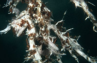
– Enlarged view – |
| • references | |
| Agerer R (1998) Tsugaerhiza tripigmentata. In Agerer R (ed) Colour Atlas of Ectomycorrhizae, plate 140, Einhorn-Verlag, Schwäbisch Gmünd. Agerer R, Trappe JM (1997) "Tsugaerhiza tripigmentatat" + Tsuga heterophylla (Rafin.) Sarg. Descr Ectomyc 2: 85-89. |
|
| • length | |
| 0 mm | Lower value of unspecified range (could be µ-s.d., but not known) |
| 20 mm | Upper value of unspecified range (could be µ+s.d., but not known) |
| • ramification presence-type | |
| monopodial-pinnate | |
| or | monopodial-pyramidal |
| • tips {per 10 mm} number | |
| 1 | Lower value of unspecified range (could be µ-s.d., but not known) |
| 3 | Upper value of unspecified range (could be µ+s.d., but not known) |
| • ramification orders | |
| 0 | Lower value of unspecified range (could be µ-s.d., but not known) |
| 1 | Upper value of unspecified range (could be µ+s.d., but not known) |
| • abundance | |
| abundant, dense | |
| • main axis diameter | |
| 0.45 mm | Lower value of unspecified range (could be µ-s.d., but not known) |
| 0.6 mm | Upper value of unspecified range (could be µ+s.d., but not known) |
| • rhizomorphs as stout, short, conical structures presence-abundance | |
| absent | |
| • rhizomorphs as short mycorrhiza-like outgrowths with blunt tips presence | |
| absent | |
| • rhizomorphs presence | |
| present | |
| • rhizomorphs frequency | |
| abundant | |
| • shape | |
| sinuous | |
| or | tortuous |
| • shape {of distal end} | |
| not inflated, cylindric | |
| • length | |
| 0 mm | Lower value of unspecified range (could be µ-s.d., but not known) |
| 6.6 mm | Upper value of unspecified range (could be µ+s.d., but not known) |
| 8 mm | Maximum value |
| • diameter | |
| 0.33 mm | Lower value of unspecified range (could be µ-s.d., but not known) |
| 0.45 mm | Upper value of unspecified range (could be µ+s.d., but not known) |
| • colour | |
| lilac, light reddish blue | |
| • older parts colour | |
| brown | |
| • mantle cortical cells visibility | |
| not visible | |
| • mantle {distinct} surface visibility | |
| present | |
| • mantle transparency | |
| not transparent | |
| • mantle laticifers visibility | |
| absent | |
| • mantle dots presence-colour | |
| absent | |
| • mantle carbonizing presence | |
| absent | |
| • mantle surface {in general} habit | |
| silvery | |
| • mantle surface {in detail} kind | |
| loosely stringy | |
| • emanating hyphae presence | |
| present | |
| • emanating hyphae abundance | |
| infrequent | |
| or | abundant |
| • cross-section shape | |
| flat | |
| • colour | |
| lilac, light reddish blue | |
| • ramification kind-frequency | |
| repeatedly into smaller filaments | |
| • connection to mantle kind | |
| oblique | |
| • origin location | |
| not specific | |
| or | distal |
| or | proximal |
| • margin habit | |
| hairy | |
| • presence | |
| absent | |
| • emanating elements presence-type | |
| rhizomorphs | |
| • blue granules presence | |
| absent | |
| • presence | |
| absent | |
| • matrix presence | |
| present | |
| • matrix location | |
| inner mantle layer | |
| • organisation | |
| plectenchymatous | |
| • mantle type | |
| hyphae rather irregularly arranged and no special pattern discernible (type B) | |
| • hyphal system kind | |
| undifferentiated | |
| • septa thickness {relative to cell walls} | |
| as thick as walls | |
| • cell shape | |
| cylindric, constricted at septa | |
| • cell pigment location-colour | |
| absent | |
| and | membranaceously yellowish |
| and | membranaceously brownish |
| and | plasmatically yellowish |
| and | vacuolarly lilac, light reddish blue |
| • cell contents presence-kind | |
| {in some cells} homogeneous, brownish | |
| • cell diameter | |
| 2 µm | Minimum value |
| 3 µm | Lower value of unspecified range (could be µ-s.d., but not known) |
| 3.5 µm | Upper value of unspecified range (could be µ+s.d., but not known) |
| 4 µm | Maximum value |
| • cell length | |
| 55 µm | Lower value of unspecified range (could be µ-s.d., but not known) |
| 100 µm | Upper value of unspecified range (could be µ+s.d., but not known) |
| • cell wall thickness | |
| 0.2 µm | Lower value of unspecified range (could be µ-s.d., but not known) |
| 0.5 µm | Upper value of unspecified range (could be µ+s.d., but not known) |
| • cell wall surface habit | |
| smooth | |
| • drops of exuded pigment presence | |
| present | |
| • organisation | |
| plectenchymatous | |
| • hyphae arrangement | |
| plectenchymatous, without pattern | |
| • cell pigment location-colour | |
| colourless | |
| or | membranaceously yellowish |
| or | membranaceously brownish |
| • cell contents presence-kind | |
| absent | |
| • cell wall thickness | |
| 0.2 µm | Mean (= average) |
| • cell wall surface habit | |
| smooth | |
| • organisation | |
| plectenchymatous | |
| • matrix kind | |
| not gelatinous | |
| • hyphae arrangement | |
| without pattern | |
| or | ring-like |
| • septa clamps presence | |
| absent | |
| • cell pigment location-colour | |
| absent | |
| or | membranaceously yellowish |
| • cell diameter | |
| 3.5 µm | Minimum value |
| 5 µm | Lower value of unspecified range (could be µ-s.d., but not known) |
| 8 µm | Upper value of unspecified range (could be µ+s.d., but not known) |
| 10 µm | Maximum value |
| • cell length | |
| 8 µm | Lower value of unspecified range (could be µ-s.d., but not known) |
| 90 µm | Upper value of unspecified range (could be µ+s.d., but not known) |
| • cell contents presence-kind | |
| absent | |
| • anatomy mantle outer mantle layer {of ectomycorrhizal tip} organisation | |
| like other parts of mantle | |
| • anatomy mantle outer mantle layer {of ectomycorrhizal tip} hyphae diameter | |
| 2.5 µm | Lower value of unspecified range (could be µ-s.d., but not known) |
| 3.5 µm | Upper value of unspecified range (could be µ+s.d., but not known) |
| • mantle thickness {apart from tip} | |
| 20 µm | Minimum value |
| 30 µm | Lower value of unspecified range (could be µ-s.d., but not known) |
| 40 µm | Upper value of unspecified range (could be µ+s.d., but not known) |
| • mantle thickness {at ectomycorrhizal tip} | |
| 15 µm | Lower value of unspecified range (could be µ-s.d., but not known) |
| 25 µm | Upper value of unspecified range (could be µ+s.d., but not known) |
| • mantle different layers presence | |
| not discernable | |
| • outer mantle layer organisation | |
| plectenchymatous | |
| • middle mantle layer organisation | |
| plectenchymatous | |
| • inner mantle layer organisation | |
| plectenchymatous | |
| • unlayered mantle hyphae tangentially length | |
| 3 µm | Lower value of unspecified range (could be µ-s.d., but not known) |
| 25 µm | Upper value of unspecified range (could be µ+s.d., but not known) |
| 40 µm | Maximum value |
| • unlayered mantle hyphae radially diameter | |
| 2 µm | Lower value of unspecified range (could be µ-s.d., but not known) |
| 4 µm | Upper value of unspecified range (could be µ+s.d., but not known) |
| • presence | |
| present | |
| • rows number | |
| 0 | Lower value of unspecified range (could be µ-s.d., but not known) |
| 1 | Upper value of unspecified range (could be µ+s.d., but not known) |
| • shape | |
| tangentially-oval, -elliptic or -cylindrical, and oriented in parallel to root axis | |
| • tangentially length | |
| 100 µm | Lower value of unspecified range (could be µ-s.d., but not known) |
| 155 µm | Upper value of unspecified range (could be µ+s.d., but not known) |
| • radially diameter | |
| 4 µm | Lower value of unspecified range (could be µ-s.d., but not known) |
| 10 µm | Upper value of unspecified range (could be µ+s.d., but not known) |
| • mean tangenial length TCt | |
| 121 µm | Mean (= average) |
| • mean shape-ratio TCq | |
| 16.6 | Mean (= average) |
| • anatomy mantle longitudinal section cortical (epidermal) cells shape | |
| tangentially-oval to -elliptic or -cylindrical, and oriented in parallel to root axis | |
| • anatomy mantle longitudinal section cortical (epidermal) cells tangentially length | |
| 83 µm | Minimum value |
| 115 µm | Lower value of unspecified range (could be µ-s.d., but not known) |
| 250 µm | Upper value of unspecified range (could be µ+s.d., but not known) |
| 320 µm | Maximum value |
| • anatomy mantle longitudinal section cortical (epidermal) cells radially diameter | |
| 12 µm | Minimum value |
| 17 µm | Lower value of unspecified range (could be µ-s.d., but not known) |
| 28 µm | Upper value of unspecified range (could be µ+s.d., but not known) |
| • anatomy mantle longitudinal section cortical (epidermal) cells mean tangential length CCt (ECt) | |
| 176 µm | Mean (= average) |
| • anatomy mantle longitudinal section cortical (epidermal) cells mean shape-ratio CCq (ECq) | |
| 8.8 | Mean (= average) |
| • presence | |
| present | |
| • kind | |
| one or half a row of cortical cells adjoining endodermis free of Hartig net | |
| • anatomy mantle longitudinal section hyphal cells around tannin cells shape | |
| roundish | |
| or | cylindrical |
| • anatomy mantle longitudinal section hyphal cells around tannin cells thickness | |
| 3 µm | Lower value of unspecified range (could be µ-s.d., but not known) |
| 4.5 µm | Upper value of unspecified range (could be µ+s.d., but not known) |
| 6 µm | Maximum value |
| • anatomy mantle longitudinal section hyphal rows around tannin cells number | |
| one | |
| • anatomy mantle longitudinal section hyphal cells around cortical (epidermal) cells shape | |
| roundish | |
| or | cylindrical |
| • anatomy mantle longitudinal section hyphal cells around cortical cells (epidermal) thickness | |
| 2 µm | Lower value of unspecified range (could be µ-s.d., but not known) |
| 2.5 µm | Upper value of unspecified range (could be µ+s.d., but not known) |
| 3.5 µm | Maximum value |
| • anatomy mantle longitudinal section hyphal rows around cortical (epidermal) cells number | |
| one | |
| • structure {in plan view} | |
| of palmetti type | |
| • lobes width | |
| 0.5 µm | Minimum value |
| 1 µm | Lower value of unspecified range (could be µ-s.d., but not known) |
| 2.5 µm | Upper value of unspecified range (could be µ+s.d., but not known) |
| 4 µm | Maximum value |
| • mantle different layers presence | |
| not discernible | |
| • outer mantle layer organisation | |
| plectenchymatous | |
| • middle mantle layer organisation | |
| plectenchymatous | |
| • inner mantle layer organisation | |
| plectenchymatous | |
| • unlayered mantle hyphae tangentially length | |
| 2 µm | Lower value of unspecified range (could be µ-s.d., but not known) |
| 20 µm | Upper value of unspecified range (could be µ+s.d., but not known) |
| 30 µm | Maximum value |
| • unlayered mantle hyphae radially diameter | |
| 2 µm | Lower value of unspecified range (could be µ-s.d., but not known) |
| 4 µm | Upper value of unspecified range (could be µ+s.d., but not known) |
| 6 µm | Maximum value |
| • presence | |
| present | |
| • rows number | |
| 1 | Minimum value |
| 2 | Mean (= average) |
| 3 | Maximum value |
| • shape | |
| tangentially-oval to tangentially-elliptic | |
| • anatomy mantle cross-section cortical (epidermal) cells shape | |
| round | |
| or | tangentially-oval to tangentially-elliptic |
| • presence | |
| present | |
| • kind | |
| one or half a row of cortical cells adjoining endodermis free of Hartig net | |
| • septal pores configuration | |
| dolipore-like structures | |
| or | globular thickenings |
| • backwards-oriented ramifications presence | |
| present | |
| • anastomoses type | |
| open, with a short bridge or bridge almost lacking | |
| • anastomoses cell wall thickness {relative to remaining cell walls} | |
| as thick as | |
| • anastomoses anastomosal bridge thickness {relative to hyphae} | |
| as thick as | |
| • anastomoses location | |
| not specified | |
| • shape | |
| not striking | |
| • drops of exuded pigment presence | |
| absent | |
| • presence | |
| present | |
| • kind | |
| acute | |
| • distance {from septum} | |
| adjacent to septum | |
| • side-branches at septum number | |
| one side-branch at septum | |
| • clamps presence | |
| present | |
| • clamps width {relative to hypha in dorsal view} | |
| thinner than | |
| • clamps outline {in lateral view} | |
| as a semicircle | |
| or | more than a semicircle |
| or | not constricted at contact point to subtending hyphal cell |
| • clamps width {relative to hypha in lateral view} | |
| as broad as | |
| or | broader than |
| • clamps hole presence | |
| absent | |
| or | present |
| • clamps blister-like structure {at basis} presence | |
| absent | |
| • presence | |
| absent | |
| • type | |
| differentiated; some hyphae very thick, which appear randomly distributed, pores of septa somtimes enlarged (type D) |
|
| • nodia presence | |
| absent | |
| • internal nodia presence | |
| absent | |
| • gelatinous matrix presence | |
| absent | |
| • gelatinized hyphae presence | |
| absent | |
| • cup-like structures on surface presence | |
| absent | |
| • conical young side-branches presence | |
| absent | |
| • a "ball" of intertwined, ramified, thin hyphae presence | |
| absent | |
| • contents presence-kind | |
| filled with brownish or yellowish substances | |
| • ampullate, trumpet-like inflated presence | |
| absent | |
| or | present |
| • ampullate inflations thickness | |
| 7.5 µm | Lower value of unspecified range (could be µ-s.d., but not known) |
| 13 µm | Upper value of unspecified range (could be µ+s.d., but not known) |
| • anatomy emanating elements rhizomorphs hyphae contents presence-kind | |
| filled with brownish or yellowish substances | |
| • anatomy emanating elements rhizomorphs hyphae ampullate, trumpet-like inflated presence | |
| absent | |
| or | present |
| • anatomy emanating elements rhizomorphs hyphae ampullate inflations thickness | |
| 7.5 µm | Lower value of unspecified range (could be µ-s.d., but not known) |
| 13 µm | Upper value of unspecified range (could be µ+s.d., but not known) |
| • presence | |
| absent | |
| • {of ectomycorrhiza former} presence | |
| absent | |
| • {of foreign origin} presence | |
| absent | |
| • mantle in section UV-filter 340-380 nm presence | |
| present | |
| • mantle in section blue-filter, 450-490 nm presence | |
| present | |
| • mantle in section green-filter, 530-560 nm presence | |
| present | |
| • geographic occurrence continent | |
| North America | |
| • plant family | |
| Pinaceae | |
| • plant genus | |
| Tsuga | |
| • plant habitat kind | |
| forests, woods | |
| • public notes | |
| Mycorrhizal systems frequently irregularly ramified; inner mantle layer in plan view at most in outlines ring-like; outer mantle layer of very tip with frequently hyphae filled homogeneously with yellowish-brown substances; emanating hyphae vacuolarly lilac; rhizomorphs with very infrequent ampullate hyphae; autofluorescence of mantle in section with UV-filter slightly blue, with blue-filter slightly yellowish green, with green-filter slightly reddish; mantle in sulfo-vanillin with some hyphae showing reddish brown contents, in KOH distinctly yellowish brown, in guaiac blue, in FeSO4 distinctly green, in lactic acid vacuolar pigment dissolving, in brillant-cresyl-blue cell walls slightly greenish, in cotton-blue cell walls slightly bluish. | |