|
|
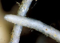
– Enlarged view – |
| • references | |
| Agerer R (1994) Pseudotomentella tristis (Thelephoraceae). Eine Analyse von Fruchtkörper und Ektomykorrhizen. Z Mykol 60: 143-158. Agerer R (1994) Pseudotomentella tristis. In Agerer R (ed) Colour Atlas of Ectomycorrhizae, plate 84, Einhorn-Verlag, Schwäbisch Gmünd. |
|
| • length | |
| 0 mm | Lower value of unspecified range (could be µ-s.d., but not known) |
| 6 mm | Upper value of unspecified range (could be µ+s.d., but not known) |
| • ramification presence-type | |
| monopodial-pyramidal | |
| • ramification orders | |
| 0 | Lower value of unspecified range (could be µ-s.d., but not known) |
| 1 | Upper value of unspecified range (could be µ+s.d., but not known) |
| • abundance | |
| solitary or in small numbers | |
| • main axis diameter | |
| 0.25 mm | Mean (= average) |
| • rhizomorphs as stout, short, conical structures presence-abundance | |
| absent | |
| • rhizomorphs as short mycorrhiza-like outgrowths with blunt tips presence | |
| absent | |
| • rhizomorphs presence | |
| absent | |
| • exploration type | |
| short distance | |
| • shape | |
| sinuous | |
| or | tortuous |
| • shape {of distal end} | |
| not inflated, cylindric | |
| or | tapering |
| • length | |
| 0 mm | Lower value of unspecified range (could be µ-s.d., but not known) |
| 3 mm | Upper value of unspecified range (could be µ+s.d., but not known) |
| 3.6 mm | Maximum value |
| • diameter | |
| 0.15 mm | Lower value of unspecified range (could be µ-s.d., but not known) |
| 0.25 mm | Upper value of unspecified range (could be µ+s.d., but not known) |
| • colour | |
| ochre, yellowish brown | |
| or | blue |
| or | white |
| • very tip colour | |
| bluish | |
| or | whitish |
| • older parts colour | |
| brown | |
| • mantle cortical cells visibility | |
| not visible | |
| • mantle {distinct} surface visibility | |
| present | |
| • mantle transparency | |
| not transparent | |
| • mantle laticifers visibility | |
| absent | |
| • mantle dots presence-colour | |
| absent | |
| • mantle carbonizing presence | |
| absent | |
| • mantle surface {in general} habit | |
| shiny | |
| or | smooth |
| • emanating hyphae presence | |
| present | |
| • emanating hyphae abundance | |
| infrequent | |
| • emanating hyphae distribution | |
| not specifically distributed | |
| or | concentrated proximally |
| • presence | |
| absent | |
| • presence | |
| absent | |
| • matrix presence | |
| present | |
| • matrix location | |
| outer mantle layer {apart from tip} | |
| or | outer mantle layer {of ectomycorrhizal tip} |
| or | middle mantle layer |
| or | inner mantle layer |
| • organisation | |
| plectenchymatous | |
| • mantle type | |
| hyphae rather irregularly arranged and no special pattern discernible (type B) | |
| and | gelatinous matrix between the hyphae (type C) |
| • matrix kind | |
| gelatinous | |
| • hyphal system kind | |
| undifferentiated | |
| and | with broad streaks of parallel hyphae |
| • septa thickness {relative to cell walls} | |
| as thick as walls | |
| and | thicker than walls |
| • septa clamps presence | |
| absent | |
| • cell shape | |
| cylindric, not constricted at septa | |
| and | cylindric, constricted at septa |
| • cell pigment location-colour | |
| membranaceously bluish | |
| • cell contents presence-kind | |
| absent | |
| • cell diameter | |
| 1.5 µm | Lower value of unspecified range (could be µ-s.d., but not known) |
| 2.5 µm | Upper value of unspecified range (could be µ+s.d., but not known) |
| • cell wall thickness | |
| 0.5 µm | Mean (= average) |
| • cell wall surface habit | |
| smooth | |
| and | with few soil particles |
| • drops of exuded pigment presence | |
| absent | |
| • organisation | |
| plectenchymatous | |
| • matrix kind | |
| gelatinous | |
| • hyphae arrangement | |
| plectenchymatous, star-like | |
| or | plectenchymatous, with broad streaks of parallel hyphae |
| • cell pigment location-colour | |
| membranaceously bluish | |
| • cell diameter | |
| 2 µm | Lower value of unspecified range (could be µ-s.d., but not known) |
| 3 µm | Upper value of unspecified range (could be µ+s.d., but not known) |
| 5 µm | Maximum value |
| • cell length | |
| 10 µm | Lower value of unspecified range (could be µ-s.d., but not known) |
| 30 µm | Upper value of unspecified range (could be µ+s.d., but not known) |
| 50 µm | Maximum value |
| • cell contents presence-kind | |
| absent | |
| • cell wall thickness | |
| 0.5 µm | Lower value of unspecified range (could be µ-s.d., but not known) |
| 1 µm | Upper value of unspecified range (could be µ+s.d., but not known) |
| • cell wall surface habit | |
| smooth | |
| • organisation | |
| plectenchymatous with pseudoparenchymatous nests of cells | |
| • matrix kind | |
| not gelatinous | |
| • hyphae arrangement | |
| without pattern | |
| • septa clamps presence | |
| absent | |
| • cell pigment location-colour | |
| absent | |
| or | membranaceously bluish |
| • cell diameter | |
| 2 µm | Lower value of unspecified range (could be µ-s.d., but not known) |
| 3 µm | Upper value of unspecified range (could be µ+s.d., but not known) |
| 5 µm | Maximum value |
| • cell length | |
| 3 µm | Lower value of unspecified range (could be µ-s.d., but not known) |
| 50 µm | Upper value of unspecified range (could be µ+s.d., but not known) |
| • cell contents presence-kind | |
| absent | |
| • anatomy mantle outer mantle layer {of ectomycorrhizal tip} organisation | |
| like other parts of mantle | |
| • anatomy mantle outer mantle layer {of ectomycorrhizal tip} matrix kind | |
| gelatinous | |
| • anatomy mantle outer mantle layer {of ectomycorrhizal tip} hyphae diameter | |
| 2 µm | Lower value of unspecified range (could be µ-s.d., but not known) |
| 3.5 µm | Upper value of unspecified range (could be µ+s.d., but not known) |
| 5 µm | Maximum value |
| • mantle thickness {apart from tip} | |
| 5 µm | Minimum value |
| 10 µm | Lower value of unspecified range (could be µ-s.d., but not known) |
| 15 µm | Upper value of unspecified range (could be µ+s.d., but not known) |
| 20 µm | Maximum value |
| • mantle thickness {at ectomycorrhizal tip} | |
| 5 µm | Lower value of unspecified range (could be µ-s.d., but not known) |
| 10 µm | Upper value of unspecified range (could be µ+s.d., but not known) |
| • mantle different layers presence | |
| not discernable | |
| • outer mantle layer organisation | |
| plectenchymatous | |
| • middle mantle layer organisation | |
| plectenchymatous | |
| • inner mantle layer organisation | |
| plectenchymatous | |
| • unlayered mantle hyphae tangentially length | |
| 2 µm | Lower value of unspecified range (could be µ-s.d., but not known) |
| 20 µm | Upper value of unspecified range (could be µ+s.d., but not known) |
| • unlayered mantle hyphae radially diameter | |
| 1.5 µm | Lower value of unspecified range (could be µ-s.d., but not known) |
| 3.5 µm | Upper value of unspecified range (could be µ+s.d., but not known) |
| • presence | |
| absent | |
| • anatomy mantle longitudinal section cortical (epidermal) cells shape | |
| rectangular | |
| or | radially-oval to -elliptic, oriented obliquely |
| • anatomy mantle longitudinal section cortical (epidermal) cells tangentially length | |
| 9 µm | Lower value of unspecified range (could be µ-s.d., but not known) |
| 20 µm | Upper value of unspecified range (could be µ+s.d., but not known) |
| 30 µm | Maximum value |
| • anatomy mantle longitudinal section cortical (epidermal) cells radially diameter | |
| 15 µm | Minimum value |
| 20 µm | Lower value of unspecified range (could be µ-s.d., but not known) |
| 45 µm | Upper value of unspecified range (could be µ+s.d., but not known) |
| 50 µm | Maximum value |
| • anatomy mantle longitudinal section cortical (epidermal) cells mean tangential length CCt (ECt) | |
| 31 µm | Mean (= average) |
| • anatomy mantle longitudinal section cortical (epidermal) cells mean shape-ratio CCq (ECq) | |
| 0.6 | Mean (= average) |
| • presence | |
| present | |
| • kind | |
| paraepidermal | |
| • anatomy mantle longitudinal section hyphal cells around cortical (epidermal) cells shape | |
| roundish | |
| • anatomy mantle longitudinal section hyphal cells around cortical cells (epidermal) thickness | |
| 1.5 µm | Lower value of unspecified range (could be µ-s.d., but not known) |
| 2.5 µm | Upper value of unspecified range (could be µ+s.d., but not known) |
| • anatomy mantle longitudinal section hyphal rows around cortical (epidermal) cells number | |
| one | |
| • structure {in plan view} | |
| of palmetti type | |
| • lobes width | |
| 1.5 µm | Minimum value |
| 2 µm | Lower value of unspecified range (could be µ-s.d., but not known) |
| 3 µm | Upper value of unspecified range (could be µ+s.d., but not known) |
| 3.5 µm | Maximum value |
| • mantle different layers presence | |
| not discernible | |
| • outer mantle layer organisation | |
| plectenchymatous | |
| • middle mantle layer organisation | |
| plectenchymatous | |
| • inner mantle layer organisation | |
| plectenchymatous | |
| • presence | |
| absent | |
| • anatomy mantle cross-section cortical (epidermal) cells shape | |
| round | |
| or | radially-oval to -elliptic |
| • anatomy mantle cross-section cortical (epidermal) cells tangentially length | |
| 5 µm | Minimum value |
| 9 µm | Lower value of unspecified range (could be µ-s.d., but not known) |
| 13 µm | Upper value of unspecified range (could be µ+s.d., but not known) |
| 19 µm | Maximum value |
| • anatomy mantle cross-section cortical (epidermal) cells radially diameter | |
| 8 µm | Minimum value |
| 12 µm | Lower value of unspecified range (could be µ-s.d., but not known) |
| 20 µm | Upper value of unspecified range (could be µ+s.d., but not known) |
| 24 µm | Maximum value |
| • anatomy mantle cross-section cortical (epidermal) cells mean tangential length CCt | |
| 10.8 µm | Mean (= average) |
| • anatomy mantle cross-section cortical (epidermal) cells mean shape-ratio CCq | |
| 0.8 | Mean (= average) |
| • presence | |
| present | |
| • kind | |
| apparently one and a half row deep | |
| • intrahyphal hyphae presence | |
| present | |
| • septal pores configuration | |
| globular thickenings | |
| • anastomoses type | |
| open, with a short bridge or bridge almost lacking | |
| • anastomoses cell wall thickness {relative to remaining cell walls} | |
| as thick as | |
| • anastomoses location | |
| not specified | |
| • shape | |
| not striking | |
| • cell pigment location-colour | |
| absent | |
| or | membranaceously brownish |
| • drops of exuded pigment presence | |
| absent | |
| • distance {from septum} | |
| adjacent to septum | |
| or | one or two hyphal diam. below the septum |
| or | in considerable distance from the septum |
| • side-branches at septum number | |
| one side-branch at septum | |
| • clamps presence | |
| absent | |
| • anatomy emanating elements emanating hyphae cell shape | |
| even | |
| or | with ellbow-like protrusions |
| • anatomy emanating elements emanating hyphae cell shape {at distal end} | |
| simple | |
| or | ramified |
| • anatomy emanating elements emanating hyphae cell diameter | |
| 3 µm | Lower value of unspecified range (could be µ-s.d., but not known) |
| 4.5 µm | Upper value of unspecified range (could be µ+s.d., but not known) |
| 6 µm | Maximum value |
| • anatomy emanating elements emanating hyphae cell wall surface habit | |
| without lens-shaped appositions | |
| or | without spindle-shaped appositions |
| or | with soil particles |
| • anatomy emanating elements emanating hyphae cell wall thickness | |
| 0.5 µm | Lower value of unspecified range (could be µ-s.d., but not known) |
| 1 µm | Upper value of unspecified range (could be µ+s.d., but not known) |
| • anatomy emanating elements emanating hyphae cell wall thickness at tip {relative to remaining cell wall} | |
| thinner | |
| or | considerably thicker, cap-like |
| • anatomy emanating elements emanating hyphae cell wall {apart from tip} evenness | |
| even in thickness | |
| or | uneven in thickness |
| • type | |
| lacking, only emanating hyphae present (type G) |
|
| • presence | |
| absent | |
| • {of ectomycorrhiza former} presence | |
| absent | |
| • {of foreign origin} presence | |
| absent | |
| • number {per cell} | |
| 2 | Mean (= average) |
| • shape | |
| round | |
| or | oval |
| • diameter | |
| 1 µm | Mean (= average) |
| • length | |
| 1 µm | Lower value of unspecified range (could be µ-s.d., but not known) |
| 1.5 µm | Upper value of unspecified range (could be µ+s.d., but not known) |
| • distance {between each other} | |
| 0 µm | Lower value of unspecified range (could be µ-s.d., but not known) |
| 4 µm | Upper value of unspecified range (could be µ+s.d., but not known) |
| • presence | |
| absent | |
| • mantle in section UV-filter 340-380 nm presence | |
| present | |
| • mantle in section blue-filter, 450-490 nm presence | |
| present | |
| • mantle in section green-filter, 530-560 nm presence | |
| present | |
| • reaction with KOH 10% presence | |
| present | |
| • reaction with lactic acid presence | |
| present | |
| • reaction with Melzer's reagent presence | |
| amyloid | |
| • amyloidity or dextrinoidity location | |
| patches of hyphal walls | |
| or | septa, globular thickenings of septa |
| • substrate | |
| in organic layer | |
| • geographic occurrence continent | |
| Europe | |
| • knowledge about association with foreign fruitbodies presence | |
| unknown | |
| • plant family | |
| Salicaceae | |
| • plant genus | |
| Salix | |
| • plant habitat kind | |
| unknown [dwarf shrubs] | |
| • family | |
| Thelephoraceae ss. Stalpers. excl. Tylospora | |
| • fruitbodies growth habit | |
| hypogeous | |
| or | corticioid |
| • public notes | |
| Autofluorescence of mantle in section with UV-filter greenish, with blue-filter yellow, with green-filter reddish; mantle in KOH with blue patches becoming olive-green, other patches brownish, in lactic acid blue patches turning to lilac-brownish. | |