|
|
|
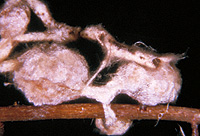
– Enlarged view – |
| • references | |
| ZAK B (1971) Characterization and classification of mycorrhizae of Douglas fir. II. Pseudotsuga menziesii + Rhizopogon vinicolor. Can J Bot 49: 1079-1084. | |
| • length | |
| 2 mm | Minimum value |
| 5 mm | Lower value of unspecified range (could be µ-s.d., but not known) |
| 15 mm | Upper value of unspecified range (could be µ+s.d., but not known) |
| 25 mm | Maximum value |
| • ramification presence-type | |
| tubercle-like | |
| • rhizomorphs presence | |
| present | |
| • rhizomorphs frequency | |
| infrequent | |
| • exploration type | |
| long distance | |
| • colour | |
| dark brown | |
| or | brown |
| or | red |
| • older parts colour | |
| black | |
| or | dark brown |
| or | lilac, light reddish blue |
| • mantle laticifers visibility | |
| absent | |
| • mantle dots presence-colour | |
| absent | |
| • mantle surface {in general} habit | |
| smooth | |
| or | not smooth |
| • diameter | |
| 0 mm | Lower value of unspecified range (could be µ-s.d., but not known) |
| 1 mm | Upper value of unspecified range (could be µ+s.d., but not known) |
| • cross-section shape | |
| round or roundish | |
| • colour | |
| concolourous to mantle | |
| • margin habit | |
| hairy | |
| • emanating elements presence-type | |
| cystidia | |
| • emanating elements cystidia location | |
| on outer mantle layer | |
| or | on rhizomorphs |
| • presence | |
| absent | |
| • organisation | |
| plectenchymatous | |
| • mantle type | |
| hyphae arranged net-like, with prominent cystidia (type D) | |
| • cell pigment location-colour | |
| membranaceously brownish | |
| • organisation | |
| plectenchymatous | |
| • cell pigment location-colour | |
| colourless | |
| • cell diameter | |
| 2 µm | Minimum value |
| 3 µm | Lower value of unspecified range (could be µ-s.d., but not known) |
| 4 µm | Upper value of unspecified range (could be µ+s.d., but not known) |
| 7 µm | Maximum value |
| • cell wall thickness | |
| 0.2 µm | Mean (= average) |
| • organisation | |
| plectenchymatous | |
| • presence | |
| present | |
| • kind | |
| protruding towards endodermis | |
| • anatomy mantle longitudinal section hyphal cells around cortical cells (epidermal) thickness | |
| 2 µm | Lower value of unspecified range (could be µ-s.d., but not known) |
| 3 µm | Upper value of unspecified range (could be µ+s.d., but not known) |
| • anatomy mantle longitudinal section hyphal rows around cortical (epidermal) cells number | |
| one | |
| • presence | |
| present | |
| • kind | |
| protruding towards endodermis | |
| • anatomy mantle cross-section hyphal cells around cortical (epidermal) cells thickness | |
| 2 µm | Lower value of unspecified range (could be µ-s.d., but not known) |
| 3 µm | Upper value of unspecified range (could be µ+s.d., but not known) |
| • anatomy mantle cross-section hyphal rows around cortical (epidermal) cells number | |
| one | |
| • type | |
| awl-shaped, bristle-like (type A) | |
| • cell wall colour | |
| brownish | |
| • presence | |
| absent | |
| • mantle in section UV-filter 340-380 nm presence | |
| present | |
| • geographic occurrence continent | |
| North America | |
| • knowledge about association with foreign fruitbodies presence | |
| unknown | |
| • plant family | |
| Pinaceae | |
| • plant genus | |
| Pseudotsuga | |
| • plant habitat kind | |
| forests, woods | |
| • family | |
| Rhizopogonaceae | |
| • fruitbodies growth habit | |
| hypogeous | |
| or | gastroid |
| • public notes | |
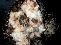
| Tubercle rind is here taken as outer mantle layer; tubercle vinaceous or of a light to darker brown, older tubercles brown-black to black with blotches of greyish bloom; inner mantle layer with labyrinthine pattern; rhizomorphs with a central core of thick hyphae; rind of tubercle (10-)25(-60) um thick; autofluorescence with UV-filter of the surface of the fresh, mature tubercles deeply velvety black and the interior is moderately bright pink to rose; in KOH hyphae turning along tears from brown to green, inner light coloured parts turned a light pink. | |