|
|
|
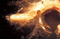
– Enlarged view – |
| • references | |
| Agerer R (1987) Studies on ectomycorrhizae V. Mycorrhizae formed by Dermocybe cinnamomea and D. sanguinea on spruce. Nova Hedwigia 44(1-2): 69-89. Agerer R (1987) Dermocybe sanguinea. In Agerer R (ed) Colour Atlas of Ectomycorrhizae, plate 8, Einhorn-Verlag, Schwäbisch Gmünd. |
|
| • ramification presence-type | |
| monopodial-pinnate | |
| or | monopodial-pyramidal |
| • rhizomorphs as stout, short, conical structures presence-abundance | |
| absent | |
| • rhizomorphs as short mycorrhiza-like outgrowths with blunt tips presence | |
| absent | |
| • rhizomorphs presence | |
| present | |
| • rhizomorphs frequency | |
| abundant | |
| • exploration type | |
| medium distance fringe | |
| • shape | |
| bent | |
| or | sinuous |
| • shape {of distal end} | |
| not inflated, cylindric | |
| • length | |
| 0 mm | Lower value of unspecified range (could be µ-s.d., but not known) |
| 2 mm | Upper value of unspecified range (could be µ+s.d., but not known) |
| 3 mm | Maximum value |
| • diameter | |
| 0.25 mm | Lower value of unspecified range (could be µ-s.d., but not known) |
| 0.35 mm | Upper value of unspecified range (could be µ+s.d., but not known) |
| 0.46 mm | Maximum value |
| • colour | |
| brown | |
| or | red |
| • very tip colour | |
| brown | |
| or | reddish |
| • older parts colour | |
| brown | |
| • mantle cortical cells visibility | |
| not visible | |
| • mantle {distinct} surface visibility | |
| present | |
| • mantle transparency | |
| not transparent | |
| • mantle laticifers visibility | |
| absent | |
| • mantle dots presence-colour | |
| absent | |
| • mantle carbonizing presence | |
| absent | |
| • mantle surface {in general} habit | |
| silvery | |
| or | not smooth |
| • mantle surface {in detail} kind | |
| densely woolly | |
| or | loosely woolly |
| • emanating hyphae presence | |
| present | |
| • emanating hyphae abundance | |
| abundant | |
| • emanating hyphae distribution | |
| not specifically distributed | |
| • cross-section shape | |
| round or roundish | |
| • colour | |
| concolourous to mantle | |
| • ramification kind-frequency | |
| repeatedly into smaller filaments | |
| • connection to mantle kind | |
| oblique | |
| • origin location | |
| not specific | |
| or | distal |
| • margin habit | |
| hairy | |
| • dimorphism presence | |
| absent | |
| • presence | |
| absent | |
| • emanating elements presence-type | |
| rhizomorphs | |
| • blue granules presence | |
| absent | |
| • presence | |
| absent | |
| • organisation | |
| plectenchymatous | |
| • mantle type | |
| ring-like arrangement of hyphal bundles (type A) | |
| • hyphal system kind | |
| undifferentiated | |
| • septa thickness {relative to cell walls} | |
| as thick as walls | |
| • cell pigment location-colour | |
| membranaceously brownish | |
| • cell diameter | |
| 4 µm | Lower value of unspecified range (could be µ-s.d., but not known) |
| 5.5 µm | Upper value of unspecified range (could be µ+s.d., but not known) |
| • cell wall thickness | |
| 0.5 µm | Mean (= average) |
| • cell wall surface habit | |
| smooth | |
| • drops of exuded pigment presence | |
| absent | |
| • organisation | |
| plectenchymatous | |
| • hyphae arrangement | |
| plectenchymatous, ring-like | |
| • cell wall thickness | |
| 1 µm | Mean (= average) |
| • organisation | |
| plectenchymatous | |
| • hyphae arrangement | |
| ring-like | |
| • cell diameter | |
| 3 µm | Lower value of unspecified range (could be µ-s.d., but not known) |
| 5 µm | Upper value of unspecified range (could be µ+s.d., but not known) |
| 6 µm | Maximum value |
| • anatomy mantle outer mantle layer {of ectomycorrhizal tip} organisation | |
| like other parts of mantle | |
| • anatomy mantle outer mantle layer {of ectomycorrhizal tip} hyphae diameter | |
| 2 µm | Lower value of unspecified range (could be µ-s.d., but not known) |
| 4 µm | Upper value of unspecified range (could be µ+s.d., but not known) |
| • mantle thickness {apart from tip} | |
| 5 µm | Minimum value |
| 10 µm | Lower value of unspecified range (could be µ-s.d., but not known) |
| 15 µm | Upper value of unspecified range (could be µ+s.d., but not known) |
| 20 µm | Maximum value |
| • mantle different layers presence | |
| not discernable | |
| or | discernable |
| • outer mantle layer organisation | |
| plectenchymatous | |
| • outer mantle layer hyphae tangentially length | |
| 3 µm | Lower value of unspecified range (could be µ-s.d., but not known) |
| 15 µm | Upper value of unspecified range (could be µ+s.d., but not known) |
| 35 µm | Maximum value |
| • outer mantle layer hyphae radially diameter | |
| 2 µm | Lower value of unspecified range (could be µ-s.d., but not known) |
| 4 µm | Upper value of unspecified range (could be µ+s.d., but not known) |
| 5 µm | Maximum value |
| • middle mantle layer organisation | |
| plectenchymatous | |
| • inner mantle layer organisation | |
| plectenchymatous | |
| • inner mantle layer hyphae tangentially length | |
| 3 µm | Lower value of unspecified range (could be µ-s.d., but not known) |
| 15 µm | Upper value of unspecified range (could be µ+s.d., but not known) |
| 35 µm | Maximum value |
| • inner mantle layer hyphae radially diameter | |
| 3 µm | Lower value of unspecified range (could be µ-s.d., but not known) |
| 6 µm | Upper value of unspecified range (could be µ+s.d., but not known) |
| • presence | |
| present | |
| • shape | |
| tangentially-oval, -elliptic or -cylindrical, and oriented in parallel to root axis | |
| • tangentially length | |
| 35 µm | Lower value of unspecified range (could be µ-s.d., but not known) |
| 63 µm | Upper value of unspecified range (could be µ+s.d., but not known) |
| 90 µm | Maximum value |
| • radially diameter | |
| 3 µm | Minimum value |
| 5 µm | Lower value of unspecified range (could be µ-s.d., but not known) |
| 13 µm | Upper value of unspecified range (could be µ+s.d., but not known) |
| 17 µm | Maximum value |
| • anatomy mantle longitudinal section cortical (epidermal) cells shape | |
| tangentially-oval to -elliptic or -cylindrical, and oriented obliquely | |
| or | tangentially-oval to -elliptic or -cylindrical, and oriented in parallel to root axis |
| • anatomy mantle longitudinal section cortical (epidermal) cells tangentially length | |
| 35 µm | Minimum value |
| 45 µm | Lower value of unspecified range (could be µ-s.d., but not known) |
| 95 µm | Upper value of unspecified range (could be µ+s.d., but not known) |
| 110 µm | Maximum value |
| • anatomy mantle longitudinal section cortical (epidermal) cells radially diameter | |
| 10 µm | Minimum value |
| 15 µm | Lower value of unspecified range (could be µ-s.d., but not known) |
| 25 µm | Upper value of unspecified range (could be µ+s.d., but not known) |
| 35 µm | Maximum value |
| • presence | |
| present | |
| • kind | |
| protruding towards endodermis | |
| or | one or half a row of cortical cells adjoining endodermis free of Hartig net |
| • mantle different layers presence | |
| not discernible | |
| or | discernible |
| • outer mantle layer organisation | |
| plectenchymatous | |
| • middle mantle layer organisation | |
| plectenchymatous | |
| • inner mantle layer organisation | |
| plectenchymatous | |
| • unlayered mantle hyphae tangentially length | |
| 3 µm | Lower value of unspecified range (could be µ-s.d., but not known) |
| 10 µm | Upper value of unspecified range (could be µ+s.d., but not known) |
| 30 µm | Maximum value |
| • unlayered mantle hyphae radially diameter | |
| 2 µm | Minimum value |
| 3 µm | Lower value of unspecified range (could be µ-s.d., but not known) |
| 5 µm | Upper value of unspecified range (could be µ+s.d., but not known) |
| 7 µm | Maximum value |
| • rows number | |
| 1 | Mean (= average) |
| 2 | Maximum value |
| • shape | |
| tangentially-oval to tangentially-elliptic | |
| • tangentially length | |
| 20 µm | Minimum value |
| 25 µm | Lower value of unspecified range (could be µ-s.d., but not known) |
| 35 µm | Upper value of unspecified range (could be µ+s.d., but not known) |
| 40 µm | Maximum value |
| • radially diameter | |
| 3 µm | Minimum value |
| 5 µm | Lower value of unspecified range (could be µ-s.d., but not known) |
| 13 µm | Upper value of unspecified range (could be µ+s.d., but not known) |
| 17 µm | Maximum value |
| • anatomy mantle cross-section cortical (epidermal) cells shape | |
| round | |
| or | tangentially-oval to tangentially-elliptic |
| • anatomy mantle cross-section cortical (epidermal) cells tangentially length | |
| 8 µm | Minimum value |
| 20 µm | Lower value of unspecified range (could be µ-s.d., but not known) |
| 35 µm | Upper value of unspecified range (could be µ+s.d., but not known) |
| 45 µm | Maximum value |
| • anatomy mantle cross-section cortical (epidermal) cells radially diameter | |
| 10 µm | Minimum value |
| 15 µm | Lower value of unspecified range (could be µ-s.d., but not known) |
| 25 µm | Upper value of unspecified range (could be µ+s.d., but not known) |
| 35 µm | Maximum value |
| • anatomy mantle cross-section hyphal cells around tannin cells shape | |
| roundish | |
| • anatomy mantle cross-section hyphal cells around tannin cells thickness | |
| 2 µm | Lower value of unspecified range (could be µ-s.d., but not known) |
| 4 µm | Upper value of unspecified range (could be µ+s.d., but not known) |
| • anatomy mantle cross-section hyphal rows around tannin cells number | |
| one | |
| • anatomy mantle cross-section hyphal cells around cortical (epidermal) cells shape | |
| roundish | |
| or | cylindrical |
| • anatomy mantle cross-section hyphal cells around cortical (epidermal) cells thickness | |
| 2 µm | Lower value of unspecified range (could be µ-s.d., but not known) |
| 3 µm | Upper value of unspecified range (could be µ+s.d., but not known) |
| • anatomy mantle cross-section hyphal rows around cortical (epidermal) cells number | |
| one | |
| • structure {in plan view} | |
| of palmetti type | |
| • intrahyphal hyphae presence | |
| absent | |
| • backwards-oriented ramifications presence | |
| absent | |
| • anastomoses type | |
| closed by a clamp, with a short bridge or bridge almost lacking (contact-clamp) | |
| • anastomoses cell wall thickness {relative to remaining cell walls} | |
| as thick as | |
| • anastomoses anastomosal bridge thickness {relative to hyphae} | |
| as thick as | |
| • anastomoses location | |
| not specified | |
| • shape | |
| not striking | |
| • cell pigment location-colour | |
| membranaceously brownish | |
| • drops of exuded pigment presence | |
| absent | |
| • clamps presence | |
| present | |
| • clamps outline {in lateral view} | |
| as a semicircle | |
| or | less than a semicircle |
| or | not constricted at contact point to subtending hyphal cell |
| • clamps width {relative to hypha in lateral view} | |
| thinner than | |
| • clamps hole presence | |
| absent | |
| • clamps blister-like structure {at basis} presence | |
| absent | |
| • presence | |
| present | |
| • abundance | |
| infrequent | |
| • distribution | |
| not specified | |
| • anatomy emanating elements emanating hyphae cell shape {at distal end} | |
| simple | |
| • anatomy emanating elements emanating hyphae cell diameter | |
| 3 µm | Lower value of unspecified range (could be µ-s.d., but not known) |
| 3.5 µm | Upper value of unspecified range (could be µ+s.d., but not known) |
| • anatomy emanating elements emanating hyphae cell wall surface habit | |
| smooth | |
| or | without lens-shaped appositions |
| or | without spindle-shaped appositions |
| • anatomy emanating elements emanating hyphae cell wall thickness | |
| 0.2 µm | Lower value of unspecified range (could be µ-s.d., but not known) |
| 0.5 µm | Upper value of unspecified range (could be µ+s.d., but not known) |
| • anatomy emanating elements emanating hyphae cell wall thickness at tip {relative to remaining cell wall} | |
| as thick as | |
| • anatomy emanating elements emanating hyphae cell wall {apart from tip} evenness | |
| even in thickness | |
| • type | |
| undifferentiated; hyphae rather loosely woven and of uniform diameter (type A) |
|
| • nodia presence | |
| absent | |
| • internal nodia presence | |
| absent | |
| • gelatinous matrix presence | |
| absent | |
| • gelatinized hyphae presence | |
| absent | |
| • cup-like structures on surface presence | |
| absent | |
| • conical young side-branches presence | |
| absent | |
| • a "ball" of intertwined, ramified, thin hyphae presence | |
| absent | |
| • ampullate, trumpet-like inflated presence | |
| absent | |
| • anatomy emanating elements rhizomorphs hyphae ampullate, trumpet-like inflated presence | |
| absent | |
| • anatomy emanating elements rhizomorphs hyphae ampullate, trumpet-like inflated presence | |
| absent | |
| • anatomy emanating elements rhizomorphs hyphae ampullate, trumpet-like inflated presence | |
| absent | |
| • anatomy emanating elements rhizomorphs hyphae ampullate, trumpet-like inflated presence | |
| absent | |
| • presence | |
| absent | |
| • {of ectomycorrhiza former} presence | |
| absent | |
| • number {per cell} | |
| 2 | Mean (= average) |
| 4 | Maximum value |
| • shape | |
| oval | |
| • geographic occurrence continent | |
| Europe | |
| • knowledge about association with foreign fruitbodies presence | |
| unknown | |
| • plant family | |
| Pinaceae | |
| • plant genus | |
| Picea | |
| • plant habitat kind | |
| forests, woods | |
| • family | |
| Cortinariaceae | |
| • subgenus-section | |
| Dermocybe sect. Sanguineae | |
| • fruitbodies growth habit | |
| epigeous | |
| or | pileate-lamellate |
| • public notes | |
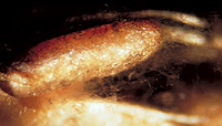
| Mycorrhizal ends reddish brown; Hartig net in plan view a weakly lobed palmetti-type; autofluorescence of mantle in section with UV-filter slightly bluish green, with blue-filter slightly green; mantle in FeSO4 slightly yellowish green, in brillant-cresyl-blue slightly greenish, in toluidin-blue violet-blue, in cotton-blue slightly blue, in acid fuchsin bownish-red. | |